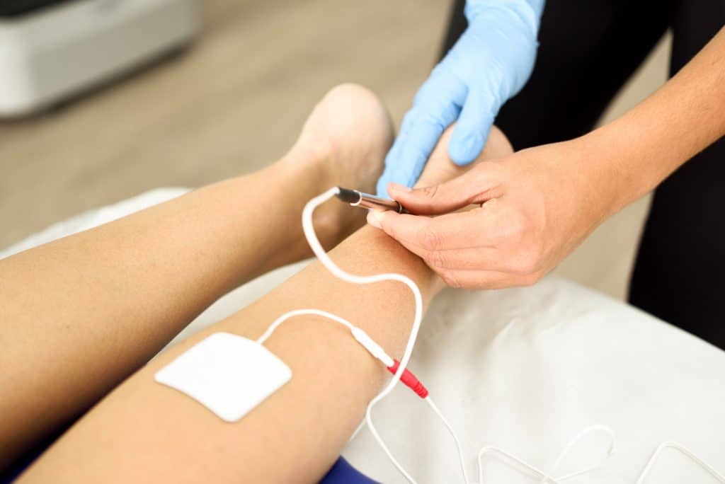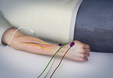

Any nerve lesion, complete or partial, from the spinal motor neurone to the intramuscular nerve branches can give rise to fibrillation. Fibrillation may persist for many months after a nerve lesion. They arise when the needle tip damages a fibre and spontaneous action potentials propagate up to the needle tip and then are extinguished.

Positive sharp waves have the same origin as fibrillation and have the same significance. Fibrillation is not visible through the skin and is an electrical sign not a clinical sign. This is detected by the EMG needle as a single fibre discharge or fibrillation (fig 1A). The effect is to make the fibre supersensitive with the result that it discharges spontaneously. Acutely denervated muscle fibres have acetylcholine receptors over the whole of the muscle fibre membrane rather than these being limited to the neuromuscular junction. Muscle fibres themselves remain viable but after a period of 7–10 days become supersensitive and fibrillations will be detectable. The sciatic nerve may be injured high along its course at the roots and plexus level underneath the pyriformis muscle in the buttock (most notoriously by injury from intramuscular injections) and along its entire course in the thigh by fracture, missile wounds orother types of injuries.SPONTANEOUS ACTIVITY Fibrillation, positive waves, and complex repetitive dischargesĪfter an acute nerve transection, nerve fibres degenerate from the site of the lesion distally. Sciatic Entrapment, Compression Or Injury Sites The stimulating electrode must be a needle electrode over the sciatic notch, which is halfway between the ischial tuberosity and greater trochanter. Place the recording electrodes on those muscles used in peroneal or posterior tibial testing. The posterior tibial nerve may be involved as part of a sciatic nerve injury at the popliteal fossa in the tarsal tunnel following ankle injury and rarely at an anterior opening of the abductor hallucis muscle. Femoral Entrapment, Compression Or Injury Sites You can also stimulate it above the inguinal ligament.īecause this nerve is difficult to stimulate in obese patients, especially above the inguinal ligament, needle electrodes may be used for that purpose. Stimulate the nerve in the groin over the femoral triangle or at Hunter’s canal. Place the active recording electrode over the vastus medialis muscle and the reference electrode on the patella.


 0 kommentar(er)
